Figure 6.
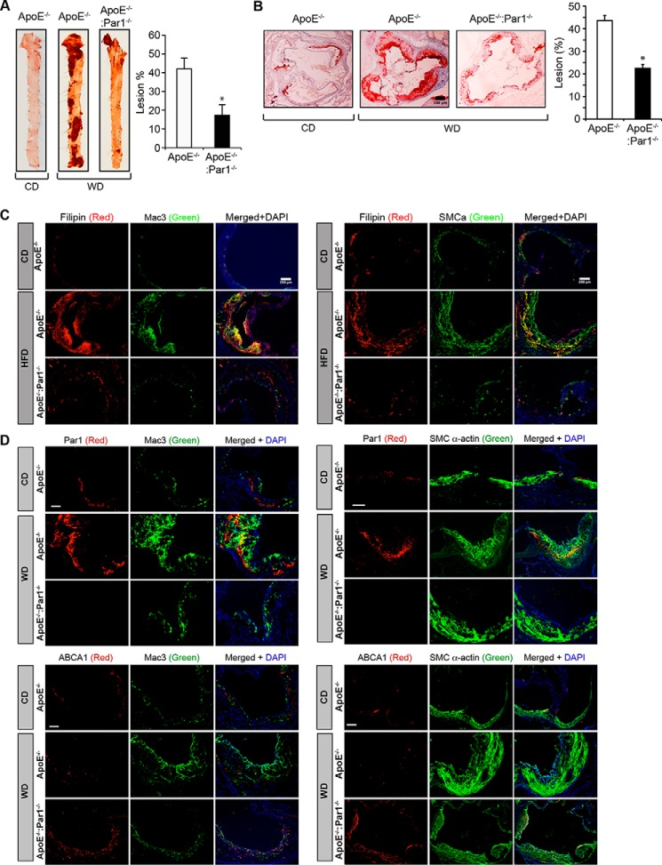
Genetic deletion of Par1 reduces atherosclerotic plaque progression. A, representative en face staining of aortas from ApoE−/− and ApoE−/−:Par1−/− mice fed with CD or WD for 16 weeks is shown, and the plaque area is presented as lesion percentage in the bar graph. B, representative Oil Red O staining of the aortic root sections of the mice described in A are shown, and the bar graph represents the quantification of the area positive for lipid staining. C and D, aortic root sections of the ApoE−/− and ApoE−/−:Par1−/− mice fed with CD or WD were stained for filipin, Par1, or ABCA1 in combination with Mac3 or SMCα-actin. Bar graphs represent mean ± S.D. (error bars) of seven animals. *, p < 0.05 versus WD-fed ApoE−/− mice. Scale bars, 100 μm (B), 200 μm (C), and 50 μm (D).

