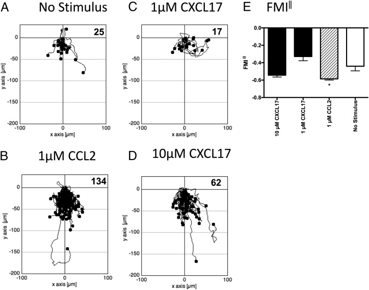
FIGURE 5.
Analysis of the migration of THP-1 cells along chemokine gradients. (A)–(D) show the paths traveled by individual cells in response to the stimuli indicated. The figures show data pooled from three experiments, with three videos analyzed per condition. Numbers of cells tracked for each condition are shown in the top right-hand corner of the panels. (E) shows the mean FMI‖ ± SEM of the data shown in (A)–(D). Statistical differences between no stimulus and the indicated stimuli were confirmed by a one-way ANOVA with Bonferroni’s multiple comparisons test. *p < 0.05.

