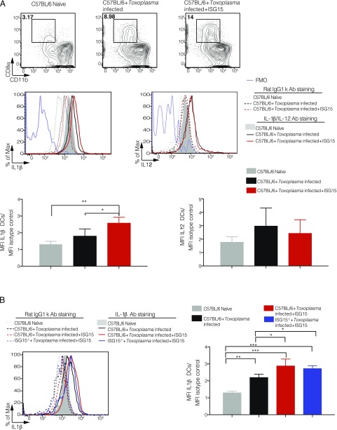
FIGURE 6.
Free ISG15 exclusively recruits IL-1β–producing CD8α+ DCs. (A) C57BL/6 mice were either infected only or infected and treated with recombinant ISG15. At day 4 p.i., cells were isolated from the peritoneal exudate, and CD8α+ population was enriched via MACS cell sorting (negative purification). IL-1β and IL-12 intracellular staining was then performed on the enriched population. Top, Representative plot of one experiment. Bottom, MFI was calculated from IL-1β and IL-12 expression in CD8α+ DCs and normalized for the isotype control for each sample. Data combined from three independent experiments. (B) C57BL/6 mice were either infected only or infected and treated with recombinant ISG15, and ISG15−/− mice were infected and treated with recombinant ISG15. Cells were isolated as in (A), and IL-1β intracellular staining was performed. Left, One representative plot. Right, MFI was calculated from IL-1β expression in CD8α+ DCs and normalized for the isotype control for each sample. Data combined from three independent experiments. Two-way ANOVA statistical analysis with Tukey test of Toxoplasma-infected and Toxoplasma-infected plus recombinant ISG15–treated mice. Only statistically significant relationships are shown. *p < 0.05, **p < 0.005, ***p < 0.0005.

