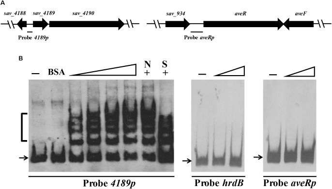
FIGURE 3.
Electrophoretic mobility shift assays (EMSAs) of SAV4189 binding to its own promoter region. (A) Schematic diagram of probes used for EMSAs. Probe 4189p: 148-bp DNA fragment, positions –145 to +3 relative to sav_4189 translational start codon. Probe aveRp: 501-bp DNA fragment, positions –476 to +25 relative to aveR translational start codon. (B) Interaction of His6-SAV4189 with probes 4189p and aveRp. Negative probe: hrdB, 118-bp DNA fragment within hrdB ORF. Negative protein control: 750 nM BSA. 0.15 nM labeled probe was added in each reaction. Concentrations of His6-SAV4189 for probes: for 4189p, 12.5, 25, 50, and 62.5 nM; for aveRp and hrdB, 125 and 250 nM. 62.5 nM His6-SAV4189 was used for competition experiments (lanes +). Lanes –: EMSAs without His6-SAV4189. Lanes N and S: competition assays with ∼100-fold excess of unlabeled non-specific competitor DNA hrdB (N) and specific probe 4189p (S). Arrows: free probes. Bracket: SAV4189-DNA complex.

