
FIGURE 3.
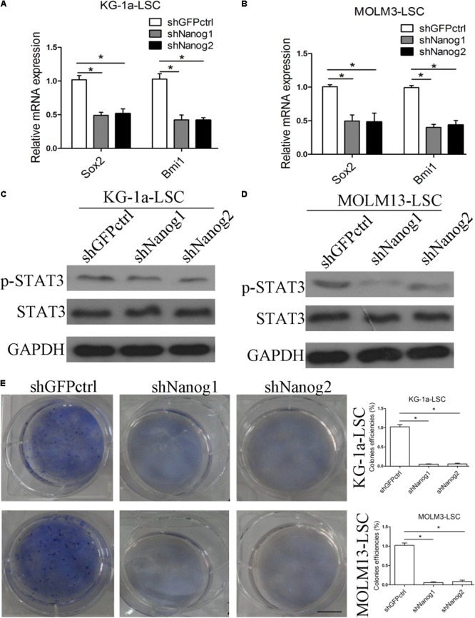
Silencing of Nanog suppresses stemness factor levels and proliferation of LSCs. (A,B) qPCR data demonstrate that the levels of Sox2 and Bmi1 was decreased. LSCs were transduced with lentiviral vector. After 72 h, LSCs were harvested. The experiments were repeated independently three times (∗P < 0.05). Data are presented as the mean ± SD. (C,D) Western blot analysis shows that p-STAT3 level was reduced. LSCs were transduced with lentiviral vectors. After 72 h, LSCs were harvested and analyzed. (E) Soft agar plates experiments demonstrate that Nanog knockdown inhibited the proliferation of LSCs. LSCs (500) were seeded in IMDM with IGF2 (20 ng/mL), EGF (20 ng/mL), bFGF (20 ng/mL), B27 supplement (1:50). After 2 weeks later, the colonies of LSCs were large enough to be visualized, and were stained with 0.5% crystal violet for 30 min at 37°C.