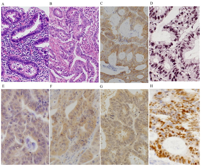
Figure 1.
Representative images of (A and B) histology and (C-H) immunohistochemical stainings in the UC-associated neoplasms. (A) The UC-associated low-grade dysplasias (Group A2) were composed of atypical columnar cells with hyperchromatic nuclei and mild nuclear stratification. (B) The UC-associated carcinomas (Group A1) showed well to moderately differentiated adenocarcinoma, invading the stroma. Group A1 lesions showed diffuse and strong cytoplasmic expressions of (C) iNOS, (E) OGG1 and (F) MTH1, and nuclear accumulations of (D) 8-OHdG and (H) p53. (G) Group A1 lesions also exhibited high cytoplasmic staining and low nuclear staining for MUTYH.

