Figure 3.

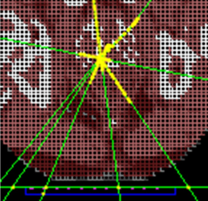
Partial axial view of voxelized patient geometry in Geant4 source position simulations. The carbon couch is shown below the patient geometry outlined in green, the Kapton substrate in blue, and the diode array in pink.

Partial axial view of voxelized patient geometry in Geant4 source position simulations. The carbon couch is shown below the patient geometry outlined in green, the Kapton substrate in blue, and the diode array in pink.