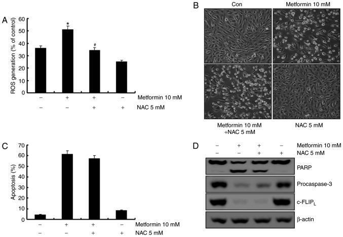
Figure 5.
Metformin-mediated apoptosis in A498 cells was not affected by reactive oxygen species generation. (A) Cells were pretreated with 5 mM NAC for 30 min and incubated with 10 mM metformin for 1 h. H2DCFDA fluorescence was quantified with flow cytometry. (B) A498 cells were incubated with 5 mM NAC or the vehicle for 30 min prior to treatment with 10 mM metformin for 24 h. Morphological changes were analyzed using a light microscope at ×200 magnification. (C) A498 cells were pretreated with 5 mM NAC or the solvent for 30 min and incubated in the presence or absence of 10 mM metformin for 24 h. The sub-G1 cell fraction was analyzed using flow cytometry. (D) A498 cells were pretreated with 5 mM NAC or the solvent for 30 min and incubated with 10 mM metformin for 24 h. PARP, procaspase-3, c-FLIPL and β-actin expression levels were analyzed using western blotting. β-actin was used as the loading control. All data are expressed as the mean ± standard deviation of three independent experiments. *P<0.05 compared with untreated cells, #P<0.05 compared with metformin-treated cells. NAC, N-acetyl-L-cysteine; PARP, poly(ADP-ribose) polymerase; c-FLIP, cellular FLICE (FADD-like IL-1β-converting enzyme)-inhibitory protein; ROS, reactive oxygen species.

