Figure 2.

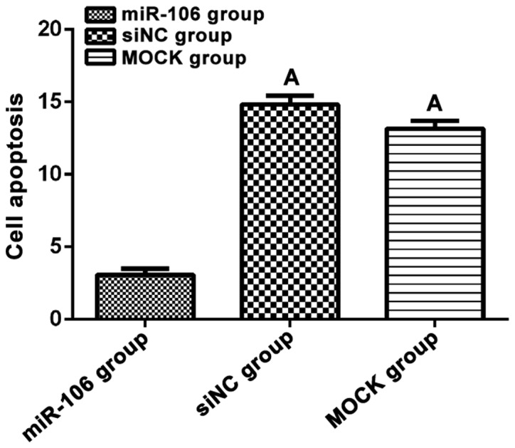
Cell apoptosis. Flow cytometry was used to detect cell apoptosis of each group after transfection. Cell apoptosis of miR-106 group was significantly lower than that in the other two groups (p<0.05). ΑP<0.05, compared with miR-106 group.

Cell apoptosis. Flow cytometry was used to detect cell apoptosis of each group after transfection. Cell apoptosis of miR-106 group was significantly lower than that in the other two groups (p<0.05). ΑP<0.05, compared with miR-106 group.