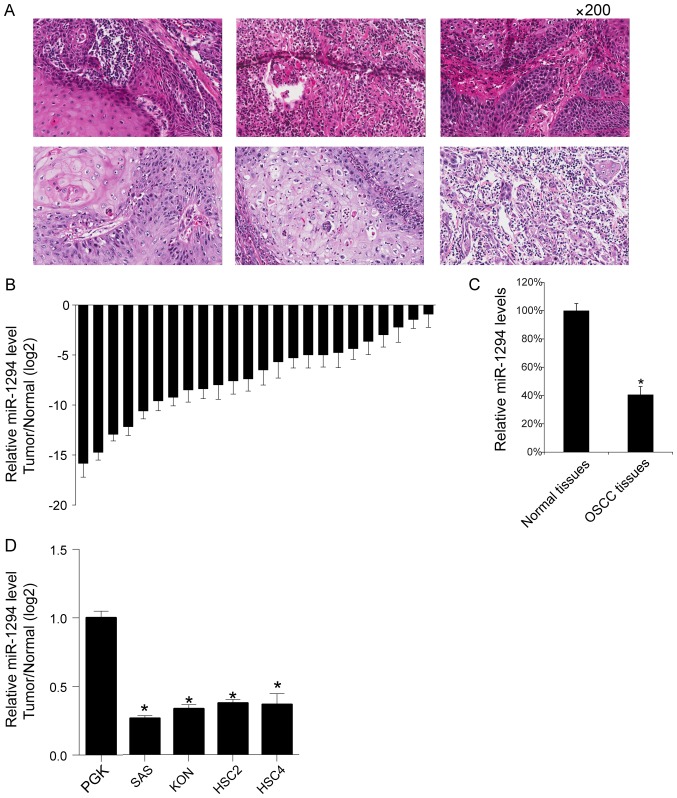
Figure 1.
Relatively low level of miR-1294 in OSCC tissue samples. (A) The representative image of OSCC tissues HE staining (magnification, ×200). The miR-1294 levels of 24 OSCC tissues samples and their matched adjacent normal tissues were assessed by qRT-PCR. Relative miR-1294 levels tumor/normal (log2) were listed (B); the mean miR-1294 values of the OSCC tissues and their matched adjacent normal tissues were also recorded. (C) *P<0.05 vs. Normal tissues. (D) The miR-545 levels of 5 OSCC cell lines (HSC2, HSC4, SAS and KON) and primary gingival keratinocytes (PGK) were assayed by qRT-PCR. *P<0.05 vs. PGK. These experiments were performed in triplicate. OSCC, oral squamous cell carcinoma.

