
Figure 1.
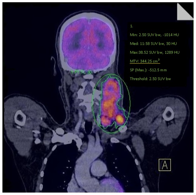
MTV assessment. MTV was measured using a SUV-based automated countering program. The margins of the tumor were drawn in order to incorporate each target lesion in the axial, coronal and sagittal 18F-FDG PET/CT images. MTV, metabolic tumor volume; SUV, standardized uptake value.