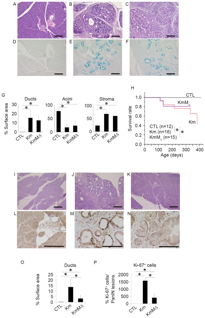
Figure 3.
c-MET expression does not influence the development of pancreatic neoplasia, and c-MET deletion in pancreatic neoplasia enhances chemosensitivity to GEM. Representative hematoxylin and eosin staining of pancreatic tissue sections from 9-month-old (A) wild-type, (B) Km and (C) KmMΔ mice. Representative images of alcian blue staining in (D) wild-type, (E) Km and (F) KmMΔ mice. (G) Quantification of fractional cross sections occupied by ductal lesions, acinar lesions or stroma in wild-type, Km and KmMΔ mice. (H) Kaplan-Meier survival rate of wild-type (n=12), Km (n=15), and KmMΔ mice (n=16). Histological analysis of the pancreas of (I) wild-type, (J) Km and (K) KmMΔ mice following 125 mg/kg GEM administration. Representative immunohistochemistry images of Ki-67 stained (L) wild-type, (M) Km and (N) KmMΔ mice. (O) Quantification of fractional cross sections occupied by ductal lesions, in wild-type, Km and KmMΔ mice. (P) Quantification of fractional cross sections occupied by Ki-67 positive lesions in wild-type, Km and KmMΔ mice. Scale bar, 100 µm. Results are presented as the mean ± standard deviation (n=5). *P<0.05. GEM, gemcitabine; CTL, control wild-type mice.

