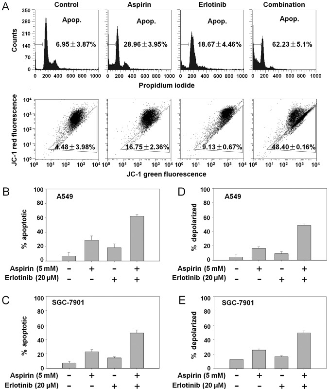
Figure 2.
Aspirin plus erlotinib induced apoptosis via mitochondrial pathway. (A) A549 cells were incubated with aspirin (5 mM), erlotinib (20 µM) or the combination for 48 h, and then cells were stained with PI (top panel)/JC-1 (bottom panel) followed by flow cytometry detection. (B and C) Cancer cells in 6-well plates were treated with drugs for 48 h and detected by flow cytometry after PI staining. (D and E) Cancer cells were treated with drugs for 48 h and detected by flow cytometry after JC-1 staining. PI, propidium iodide; JC-1, 5,5′,6,6′tetrachloro-1,1′,3,3′-tetraethylbenzimidazol-carbocyanine iodide.

