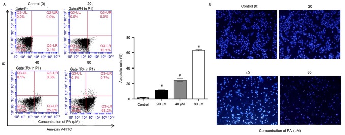
Figure 2.
Effect of PA on the apoptosis of SGC-7901 gastric cancer cells. SGC-7901 cells were treated with different concentrations of PA (20, 40 and 80 µM) for 24 h. (A) Annexin V-FITC/PI staining and (B) Hoechst 33258 fluorescence staining were used to detect cell apoptosis. Magnification, −200. #P<0.001 vs. the control group. PA, pachymic acid.

