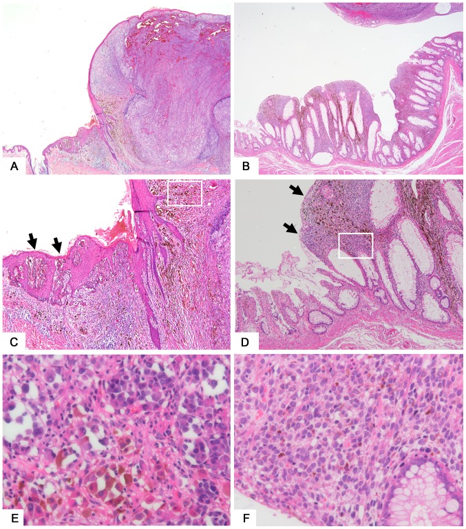
Figure 1.
Representative features of skin and gastrointestinal melanomas. (A) T4 skin melanoma tissue sample taken from the back. Magnification, −20. (B) Malignant melanoma of the rectum. Magnification, −20. (C) Melanoma in situ (arrow) seen adjacent to the nodular lesion of skin melanoma. Magnification, −100. (D) Melanoma cells (arrow) seen in the mucosal layer adjacent to the elevated lesion of gastrointestinal melanoma. Magnification, −100. (E) Magnification of a squared area of part (C). Melanoma cells showing hyperchromatic nuclei with melanin deposition in the cytoplasm. Magnification, −400. (F) Magnification of a squared area of part (D). Magnification, −400.

