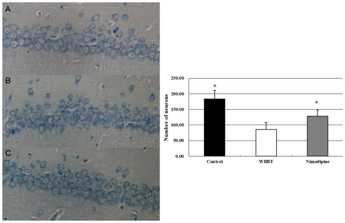
Figure 3.
Decreased numbers of neurons following WBRT. The results by Nissl staining using a microscope (magnification, ×400) revealed that the neurons in the CA1 region exhibited intact morphology in the control (A) but the neurons in the WBRT (B) appeared to be decreased and exhibited degeneration. However, nimodipine (C) group reserved more intact neurons compared with WBRT. The graph revealed that the number of neurons in hippocampal DG region in WBRT was significantly decreased compared with control (P<0.01) or nimodipine (P<0.01) group. *P<0.05 vs. WBRT. WBRT, whole brain radiotherapy; DG, dentate gyrus.

