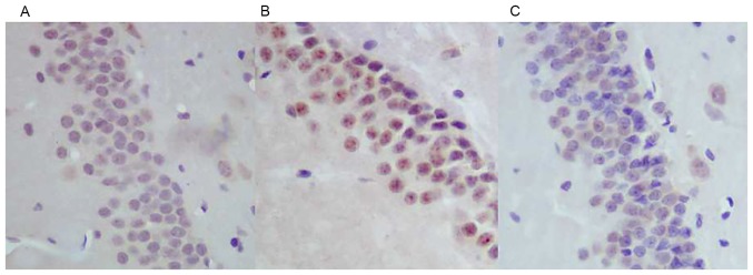
Figure 4.
Caspase-3 staining in the hippocampal DG region. The expression levels of caspase-3 in (A) control, (B) WBRT and (C) nimodipine groups. The results demonstrated that the expression of caspase-3 increased in the hippocampal CA1 region in the WBRT group compared with that in control and nimodipine groups. WBRT, whole brain radiotherapy; CA, Cornu Ammonis (magnification, ×400).

