
FIGURE 1.
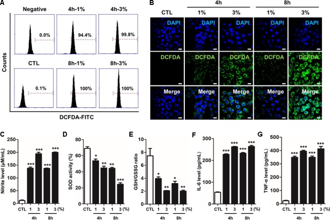
CSE-induced intracellular RONS stress and inflammation in human alveolar epithelial cells. The A549 epithelial cells were incubated in the presence (1–3%) or absence of CSE for 8 h, followed by a measurement of the intracellular redox states. (A) The fixed quantity of oxidized DCF-DA probe was evaluated by flow cytometry. The x-axis indicate the intensity of intracellular DCF-DA fluorescence, and the y-axis represent the mean number of live cells. (B) The cells exposed to CSE were loaded with DCF-DA for 30 min before being imaged using confocal laser scanning microscopy. A amount of DCF-DA is linked to live cells damaged by ROS. (C) The concentration of nitrite released into the culture medium was determined using the Griess assay 4 h or 8 h after CSE exposure. (D) Total SOD activity was measured in cell lysate exposed to CSE. (E) The ratio of glutathione (GSH)/oxidized glutathione (GSSG) was detected at different reaction time using a colorimetric assay at OD412nm. The calculated the total GSH and GSSG level of test samples based on the standard curve of ∆OD412nm/min verses GSH or GSSG standard solutions. A decreased ratio of GSH-to-GSSG is an indication of oxidative stress. The level of (F) IL-6 and (G) TNF-α were determined by ELISA in culture supernatants. The results are represented by average value of data (±SE). CTL, no treatment. ∗p < 0.05, ∗∗p < 0.01, and ∗∗∗p < 0.001 vs. CTL.