
FIGURE 2.
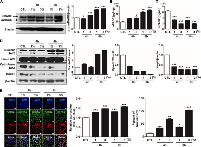
CSE-induced alternations of RAGE distribution through Nrf2 nuclear migration in human alveolar epithelial cells. The expression level of RAGE and Nrf2-related proteins was detected in A549 cells incubated in the presence (1–3%) or absence of CSE. (A) Western blotting shows the expression levels of xRAGE and mRAGE, membrane-bound RAGE isoform in cell lysates. Net intensity of RAGE was normalized to β-actin. The levels of (B) mRAGE in lung homogenates and (C) secreting sRAGE in culture supernatants of cell after CSE exposure were detected by ELISA analysis. The concentrations of proteins were calculated based on each standard curve data. The results are represented by average value of data (±SE). (D) The expression of Nrf2 migration from cytoplasm into nuclear location was determined by western blot. The Lamin A/C and β-actin were used for loading control of the nuclear protein and cytoplasmic protein, respectively. (E) The confocal microscopy analysis shows the protein expression of RAGE (green), the localization of Nrf2 (red) and merged images by immunofluorescence staining. Nuclei stained with DAPI (blue). The number of RAGE-positive cells, Nrf2-postivie cells and double-positive cells chosen fields in three randomly at ×600 magnification. The results are represented by average value of data (±SE). CTL, no treatment. ∗p < 0.05, ∗∗p < 0.01, and ∗∗∗p < 0.001 vs. CTL.