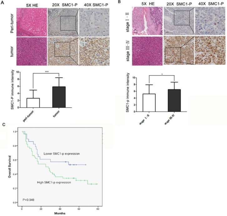
Figure 5.
SMC1 phosphorylation expression is correlated with poor prognosis in human HCC. (A) The expression level of SMC1 phosphorylation protein in HCC (n = 78) and paired peritumoral liver tissue (n = 78) was detected by immunohistochemical staining (top). Scale bars, 20 μm. Scores indicated SMC1 phosphorylation levels by a 20-point quantification scale (bottom), ***P<0.001. (B) Top, immunohistochemical staining of SMC1 phosphorylation was displayed in clinical HCC at different clinical stages. Scale bars, 20 μm. Bottom, column plot analysis of the SMC1 phosphorylation expression in HCC samples at different clinical stages, *P<0.05. (C) Kaplan-Meier survival curve showed that high SMC1 phosphorylation expression (score 5-10, n = 78) was associated with poorer overall survival compared to low SMC1 phosphorylation expression (score 0-4, n = 78), *P<0.05. Data were expressed as means ± SD. SMC1-P, SMC1 phosphorylation; WT, wild-type.

