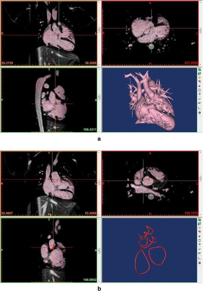
Fig. 2.

Segmentation process in a commercially available software program (Mimics®, Materialise, Leuven, Belgium) using magnetic resonance angiograms from a patient with congenitally corrected transposition of the great arteries with a ventricular septal defect. a Segmentation using thresholding and manual edition with a volume rendered image on the right lower panel. b Linear representation of the cardiac valvar attachments. A few points of attachment sites of each cardiac valve were marked and connected using a tool called “spline”
