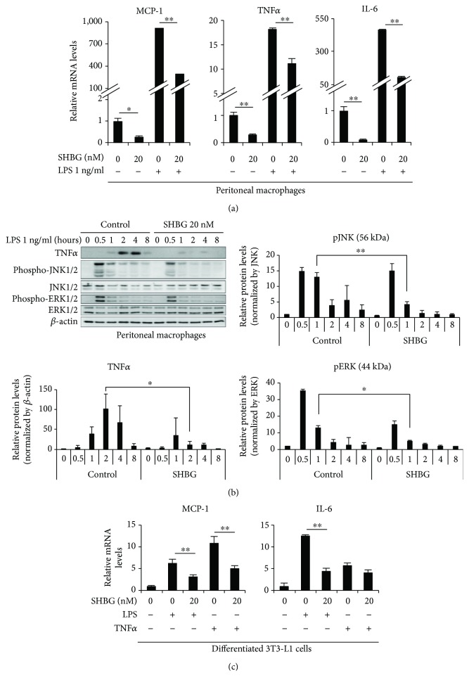
Figure 1.
SHBG inhibits inflammatory cytokine levels in peritoneal macrophages and differentiated 3T3-L1 cells. (a) Peritoneal macrophages from C57BL/6 mice were treated with SHBG overnight, followed by 1 ng/ml LPS stimulation for 8 hrs. mRNA levels of inflammatory cytokines were measured by RT-PCR. Student's t-test was performed. Data are the means ± S.D. (n = 4, ∗p < 0.05, ∗∗p < 0.01). (b) Peritoneal macrophages from C57BL/6 mice were treated with SHBG protein overnight, followed by 1 ng/ml LPS stimulation for the indicated times. Inflammatory signaling was evaluated by Western blotting. Each band was quantified using ImageJ. Relative intensities are shown. Data are the means ± S.D. (n = 3, ∗p < 0.05, ∗∗p < 0.01). (c) Differentiated 3T3-L1 cells were treated with SHBG proteins overnight, followed by 1 ng/ml LPS or 1 ng/ml TNFα stimulation for 24 hrs. mRNA levels of MCP-1 and IL-6 were measured by RT-PCR. Student's t-test was performed. Data are the means ± S.D. (n = 3, ∗∗p < 0.01).

