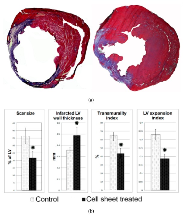
Figure 4.
Morphometric evaluation of the ventricular in rats 14 days after myocardial infarction and cell sheet therapy. (a) Representative images of Mallory-stained cross sections of myocardium. Collagen fibers are stained blue and viable tissue stained purple. (b) Histograms summarizing quantitative data: infarct size, infarct wall thickness, transmurality and left ventricular expansion indices 14 days after infarction. Data is presented as mean±SD. ∗p<0.05 versus control.

