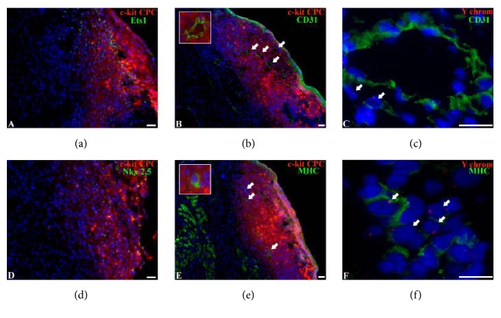
Figure 5.
Differentiation and Y-chromosome detection in CPC-based cell sheets. (a, b) Differentiation of CPC in cell sheets toward vascular lineage. Representative images of immunofluorescence staining of the heart sections with antibodies against endothelial marker Ets-1 (a) and CD31 (b). Grafted cells were identified by CM-DIL (red fluorescence). Arrows indicate CM-DIL+ vessels. (c) Y-chromosome detection in CPC-derived endothelial cell. Representative images of immunofluorescent staining of heart sections with antibodies against endothelial marker CD31 and Y-chromosome. Arrows indicate Y-chromosome+ endothelial cells. (d, e) Differentiation of CPC in cell sheets toward cardiomyocyte lineage. Representative images of immunofluorescent staining of heart sections with antibodies against cardiomyocyte markers Nkx2.5 (d) and MHC (e) (β-myosin heavy chain). Grafted cells were identified by CM-DIL (red fluorescence). Arrows indicate CM-DIL+ cardiomyocytes. (f) Y-chromosome detection in cardiomyocytes. Representative images of immunofluorescence staining of the heart sections with antibodies against cardiomyocyte marker MHC and Y-chromosome probe. Arrows indicate Y-chromosome+ cardiomyocytes.

