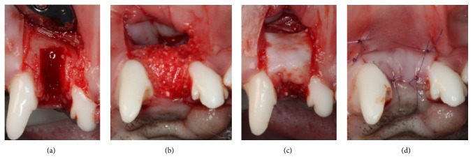
Figure 1.
Clinical photographs of the surgical procedures. (a) Extraction and defect creation (4 mm wide and 6 mm high), (b) placement of either demineralized bovine bone mineral with 10% collagen (DBBM-C) soaked with hypoxia-inducible factor 1α (HIF1A) or DBBM-C only in the defect and the upper part of the socket, (c) coverage of the defect using a collagen membrane, and (d) primary flap closure.

