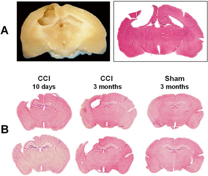
Fig. 1.

Chronic brain pathology following CCI. Representative photograph of paraformaldehyde-perfused rat brain spacemen collected at 3 months after induction of CCI and corresponding hematoxylin–eosin (H&E)-stained coronal 8-μm paraffin section. The photographs demonstrate marked brain degradation at chronic stages resulted in loss of cortical tissue (cavitation) and significant reduction in hippocampal size ipsilateral to the injury at chronic time points (a). Representative photographs of H&E-stained coronal 8-μm paraffin mouse brain sections demonstrating progression of hippocampal pathology at subacute and chronic time points after CCI. At subacute time point (10 days after CCI) the ipsilateral hippocampus is enlarged due to focal edema that is still evident at this time point, whereas at chronic time point after CCI, the ipsilateral hippocampus is reduced in size due to neurodegenerative processes resulting in neuronal death and loss of brain tissue. Cortical cavitations in CCI-injured mice are presented at both time points. In contrast, no obvious brain pathology is observed in animals at 3 months after sham injury (b)
