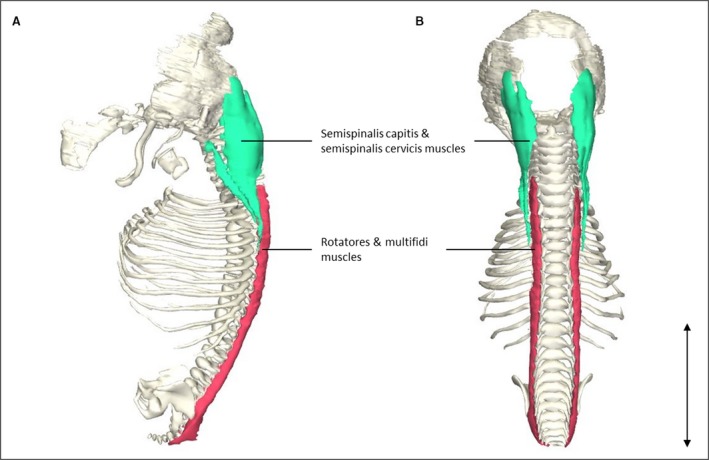
Figure 1.

Transversospinal muscle reconstrunctions. (A) Left lateral view of the head and vertebral column of a stage 23 human embryo (56–60 days of development), specimen number 950. Note that the semispinalis capitis and semispinalis cervicis muscles (green) are depicted as one structure because they were histologically inseparable. The same holds for the rotatores and multifidi muscles (red). (B) Dorsal view of the same reconstruction as in (A). The semispinalis capitis and semispinalis cervicis muscles are relatively large in size compared with the rotatores and multifidi muscles, whereas in adults the differences in size between these muscles are minimal, possibly due to a craniocaudal developmental gradient that causes craniocaudal differences in size. Scale bar: ~5 mm.
