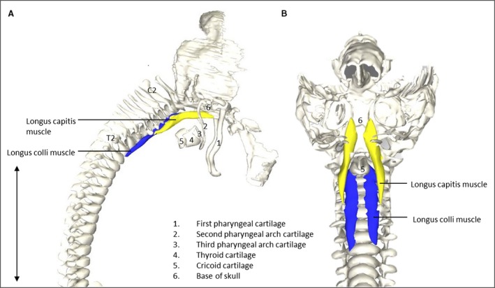
Figure 2.

Vertebrate muscles of the neck. (A) Right lateral view of the head region of a stage 23 human embryo (56–60 days of development), specimen number 950. Longus capitis (Lca, yellow) and longus colli (Lco, blue) muscles depicted on vertebrate column, head bones and neck cartilage. (B) Caudofrontal view of head and neck region of the same specimen as in (A). LCa and LCi depicted on vertebrate column, head bones and cartilage. To increase clarity, the first pharyngeal arch, hyoid and thryoid bone were excluded. Note that the relative size and shape of the muscles to the skull and vertebrae are identical, as would be expected in mature humans (Gilroy et al. 2009). Scale bar: ~5 mm.
