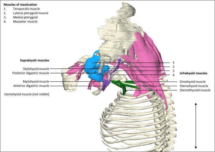
Figure 3.

Head and neck muscles. Left lateral view of the head and neck region of a stage 23 human embryo (56–60 days of development), specimen number 950. Skeletal structures are depicted in white, eye and neck musculature in pink, suprahyoid muscles in blue, infrahyoid muscles in green and muscles of mastication in purple. Suprahyoid and infrahyoid muscles show a high degree of segregation. Note that the muscles of mastication have a small size relative to the bones and surrounding muscles when compared with the relative adult sizes. Suprahyoid and infrahyoid muscles are already individually partitioned from the surrounding muscles and have the same position as in adult anatomy (Gilroy et al. 2009). The sternocleidomastoid muscles are excluded. Scale bar: ~5 mm.
