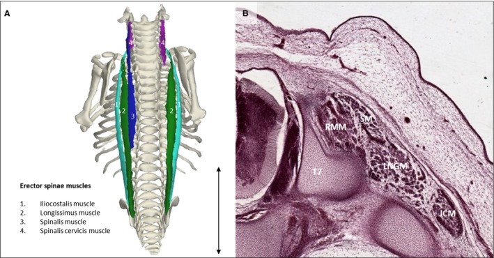
Figure 4.

Erector spinae muscles. (A) Dorsal view of the back of a stage 23 human embryo (56–60 days of development), specimen number 950. Erector spinae muscles are depicted along with the bones of the body. The different muscles are the iliocostalis muscle (light green), longissimus muscle (green), spinalis muscle (blue) and spinalis cervicis muscle (pink). On the right side the spinalis muscle is excluded, as are other muscles. (B) Transversal serial section view of the left erector spinae muscles of the same specimen as in (A) at the level of T7. Note that the erector spinae muscles have separated bodies and hence are already individually recognizable at this stage of development. LNGM, longissimus muscle; ICM, iliocostalis muscle; RMM, rotatores and multifidi muscles; SM, spinalis muscle. Scale bar: ~5 mm.
