
Figure 2.
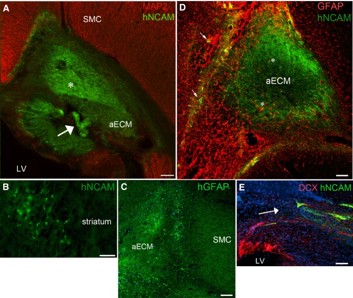
One week post grafting: (A) shows a graft of artificial ECM (aECM) containing hNCAM + cells (green) positioned deep to the host sensorimotor cortex (SMC) close to the lateral ventricle (LV). Some of the hNCAM+ cells were in a disorganised mass (asterisk), whereas some had organised themselves into a convoluted ribbon or tube (arrow). (B) Group of hNCAM + cells that have migrated away from the graft into the nearby striatum. Grafted cells also expressed hGFAP (C) and could be seen around the edges of the graft at the interface with host tissue. (D) Illustrates how host astrocytes (GFAP +, red) infiltrated the grafts (asterisks) mingling with hNCAM + grafted cells (green) in the aECM. Grafted cells also infiltrated the host tissue (arrows). (E) Shows how host DCX + cells (red), presumably newborn neuroblasts, migrated towards and mingled with hNCAM + grafted cells (green). The arrow indicates the direction of migration. Blue staining in (C) and (E) is the nuclear stain DAPI. Scale bars: (A,C,D) 200 μm; (B) 50 μm; (E) 30 μm.