
Figure 3.
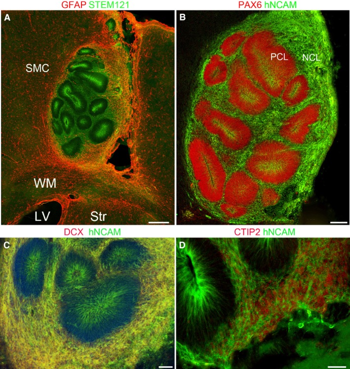
Four weeks post‐grafting. (A) Graft containing cells positive for the human stem cell marker STEM123 (green) within the host sensorimotor cortex (SMC) dorsal to the subcortical white matter (WM), lateral ventricle (LV) and striatum (Str) all positive for GFAP (red). Grafted cells are arranged in rosettes with high expression of STEM121 in cells close to the centre of the rosettes and in the looser arrangement of grafted cells surrounding the rosettes. Host astrocytes (red) have invaded the graft, particularly at the margins. (B) Dense packed layer of progenitor cells (PCL) expressing PAX6 (red) and hNCAM (green). The hNCAM was more strongly expressed in cells at the inner, luminal surface of the PCL (B–D). hNCAM was strongly expressed in the more loosely packed layer of post‐mitotic neurons (NCL; B). (C) These neurons also co‐expressed DCX and hNCAM (yellow) and some co‐expressed CTIP2 (nuclear, red) with hNCAM (green) as shown in (D). Scale bars: (A–B) 200 μm; (C) 100 μm; (D) 50 μm.