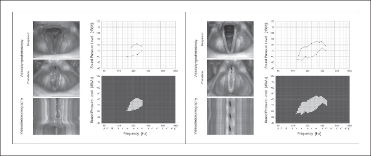
Fig. 2.

Phonomicrosurgery-induced changes of objective findings in videolaryngostroboscopy [VLS], videostrobokymography, and VRP measurements in a 52-year-old female office worker with chronic manifestation of Reinke’s edema (RE). Left: the preoperative status reveals bilateral RE (Yonekawa type III) with irregular vocal fold oscillations and absent mucosal wave propagation. The VRP envelope curves of loudest (black line) and softest singing (blue line) show small dynamic and frequency range. Polygon visualisation (green squares) depicts small VRP area, and VEM calculation results in a low value (VEM = 60). Right: three months postoperatively, the vocal folds present slim and irritation-free with a straight margin. The RE on both sides are completely removed, the healing process is finished (scar-free). The glottal closure is complete, vocal fold oscillations have normalised (mucosal wave propagation regular and symmetric). The VRP shows improved dynamic and frequency range with larger VRP area, hence VEM calculation results in a higher value (VEM = 100).
