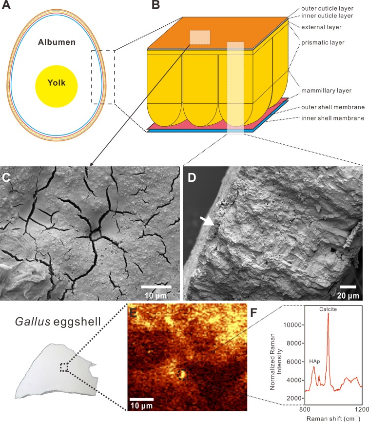
Figure 1. Cross-sectional view, SEM images, and Raman imaging and spectrum of a Gallus gallus domesticus egg and eggshell.
The Raman image and spectrum were collected using 532 nm excitation wavelength and a 50x objective with RI. (A) The generalized anatomy of an egg. (B) The chicken eggshell comprises three crystalline layers, including the mammillary layer, prismatic layer, and external layer. The cuticle layer overlying the calcareous eggshell is further divided to two layers, including a HAp inner layer and a proteinaceous outer layer. The shell membrane, namely membrane testacea, is also characterized by two layers. (C) SEM image of the cuticle on the surface of the Gallus eggshell, showing a patchy and cracked pattern. (D) SEM image of the radial section of the Gallus eggshell. The white arrow indicates the cuticle layer that lies on the calcitic eggshell. (E) Raman chemical image with peak targeting at 967 cm−1 which is attributed to HAp. The yellow area represents the patchy distribution of the inner HAp cuticle layer on the Gallus eggshell. The calcitic eggshell that is not covered by the inner HAp cuticle layer is shown in the brown area. The dotted circle corresponds to the spectrum shown in (F). (F) Spectrum collected in the dotted-circle area of the Raman chemical image shown in (E). Two significant peaks at 967 cm−1 and 1,087 cm−1 represent HAp and calcite, respectively. Credit of the SEM images and drawings: Tzu-Ruei Yang (Universität Bonn).

