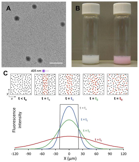
Fig. 1.
Photoactivatable fluorescent nanoparticle probes. (A) Electron micrograph of photoactivatable PLGA-PEG nanoparticles after dehydration. Scale bar is 200 nm. Fully hydrated particles in buffer solution had a diameter of 160 nm, as measured by dynamic light scattering (Table S1). (B) Photograph of particle suspensions in water before (left vial) and after (right vial) UV light exposure, demonstrating activation of the caged rhodamine. (C) Schematic of a photoactivatable nanoparticle diffusion assay (PANDA) experiment, illustrating activation of fluorescence within a rectangular region by brief 405 nm laser irradiation and subsequent diffusion of activated particles from the region. Top row: Field of view before and after activation, showing particles diffusing over time. Bottom plot: Time-dependent fluorescence intensity along the x-direction.

