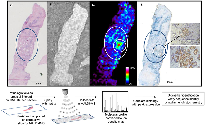
Fig. 1.

Workflow of matrix-assisted laser desorption ionization (MALDI) imaging mass spectrometry (IMS) for biomarker discovery. a a pathologist reviews the hematoxylin and eosin (H&E)-stained tissue and circles a region of prostate cancer. b a serial section is placed on a conductive slide and sprayed with matrix. c the spatial expression of the m/z 4,355 MEKK2 peptide in the prostate tissue collected using MALDI-IMS with a raster width of 200 μm correlates to the circled prostate cancer region. The relative expression of m/z 4,355 is color-coded according to the inset scale. d immunohisto-chemistry (IHC) staining of MEKK2 expression, showing strong staining in the circled areas that correspond to regions of high m/z 4,355 expression. Magnified views of the stained prostate cancer cells are shown in the insets: top ×10 and bottom ×40
