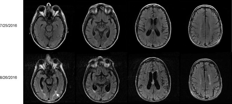
Figure 1. Serial brain MRIs.

Top panels show a normal brain structure in cross section views of T2-FLAIR scans which were obtained at the initial presentation. Repeat scans five days before the patient’s death are shown in the bottom panels. There was interval development of brain atrophy with prominence of the ventricles and sulci as well as periventricular white matter enhancement (white arrow).
