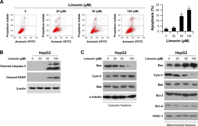
Figure 4.
Limonin induced cell apoptosis in HepG2 cells.
Notes: After limonin treatment for 24 h, HepG2 cells were subjected to Annexin V/propidium iodide double staining and FACS analysis (A) or Western blotting with indicated antibodies (B). The asterisks (*p<0.05, Student’s t-test) indicated significant difference versus the control. (C) Limonin increased Bax binding to mitochondria and induced cytochrome C release. After limonin treatment, the cytosolic and mitochondrial fractions were isolated, and the expressions of given proteins were examined with indicated antibodies.

