Figure 3.
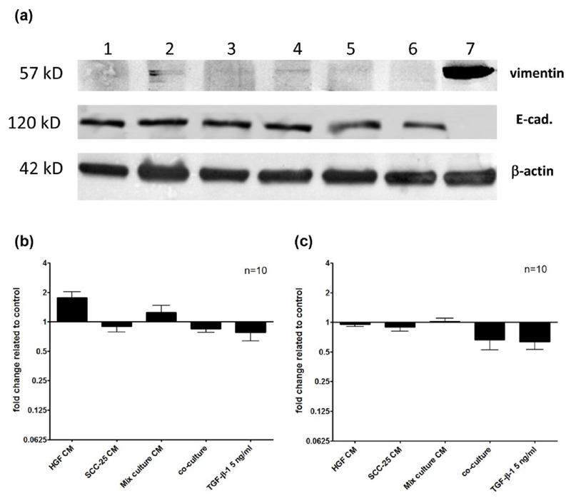
Fold change of vimentin and E-cadherin protein related to the control. The protein synthesis of vimentin, E-cadherin and β-actin in SCC-25 cells was determined using western blot analysis. β-actin was applied as a loading control. Fibroblasts (a, lane 7) served as positive control cells for vimentin, since this protein was contained in undetectable levels in parental SCC-25 cells. (a) Typical western blot of 57 kDa vimentin, 120 kDa E-cadherin, and 42 kDa β-actin in samples: (1) control SCC-25 treated with albumin medium; (2) SCC-25 treated with FIB CM; (3) SCC-25 treated with SCC-25 CM; (4) SCC-25 treated with mixed-culture CM; (5) SCC-25 co-cultured with fibroblasts; (6) SCC-25 treated with TGF-β1 (5 ng/mL); and (7) cultured fibroblasts, positive control for vimentin and negative control for E-cadherin (CM: conditioned medium). In FIB and mixed-culture CM-treated SCC-25 cells, a faint vimentin band appeared. (b–c) The band intensities were analyzed with densitometry. The different treatments were related to control cells treated with albumin-containing medium only. Densitometry graphs covered 10 comparable western blot experiments. (b) Two times upregulation of vimentin was shown by treatment with FIB CM. (c) E-cadherin showed decrease in co-culture and after TGF-β1 treatment.

