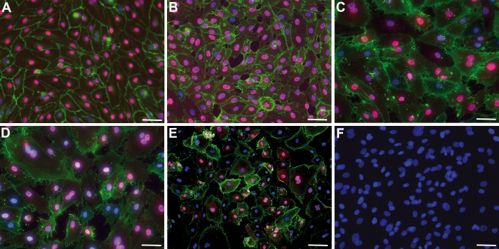
Fig 1. Culture of lymphatic endothelial cells and fibroblasts.
Immunocytology showing CD31 (green) and PROX1 (magenta). Nuclei are counter-stained with DAPI (blue). A) Foreskin-derived HD-LECc4; B) Foreskin-derived HD-LECc2; C) Patient-derived Ly-LEC-12, D) Patient-derived Ly-LEC-2; E) Patient-derived Ly-LEC-10; F) Patient-derived fibroblasts (Ly-F-12) do not express CD31 or PROX1. Bar = 45 μm in A-D,F, and 90 μm in E.

