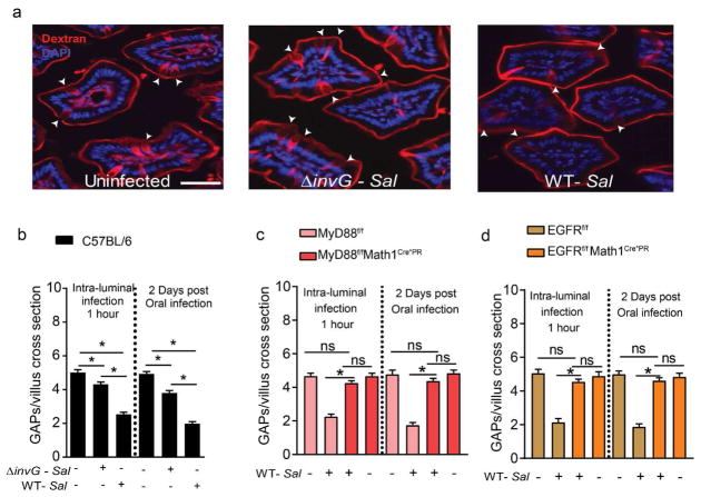
Figure 1. Goblet cell-associated antigen passage (GAP) formation is inhibited by Salmonella in a MyD88 and EGFR dependent manner.
(a) Fluorescent images of SI villus cross section of C57BL/6 mice given luminal 10kD dextran (red) and DAPI (blue), one hour after receiving luminal PBS (left), 5×10⁸ CFU invasion deficient Salmonella (ΔinvG Sal; center) or 5×10⁸ CFU wildtype Salmonella (WT-Sal; right). (b) Density of GAPs in C57BL/6 mice, given 5×10⁸ CFU or wildtype or ΔinvG Salmonella in the SI lumen 1 hr earlier (left panel) or given 5×10⁷ CFU wildtype or ΔinvG Salmonella orally 2 days earlier (right panel). (c) MyD88fl/fl Math1Cre*PR and (d) EGFRf/f Math1Cre*PR mice and littermate controls were treated with RU486 to delete MyD88 or EGFR from GCs and were administered with 5×10⁸ CFU of wildtype Salmonella or PBS for 1 hour within the SI-lumen (left panel), or gavaged with 5×10⁷ CFU wildtype Salmonella and SI tissue sections were evaluated 2 days later (right panel). Graphs depict the density of GAPs in SI villus cross section of uninfected and infected mice. Data represented as the mean ± SEM. Scale bar in a= 50 μm. *p<0.05, ns = not significant. n=5 or more mice with 60 or more villus cross sections per mouse examined for each condition.

