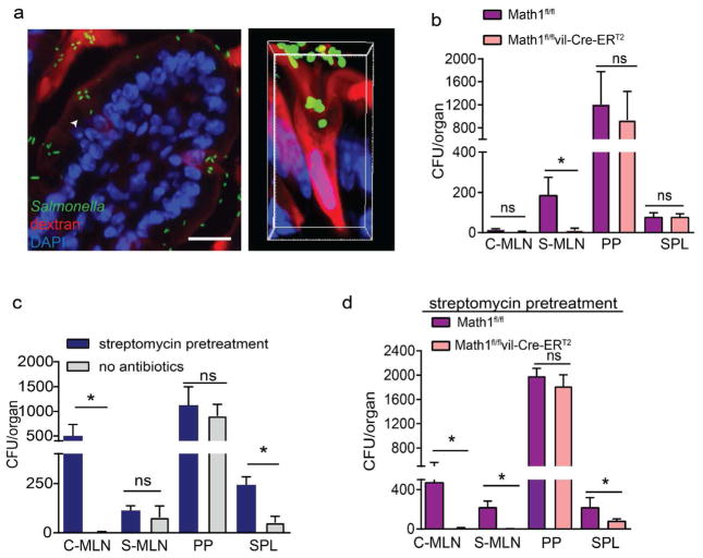
Figure 4. Dissemination of Salmonella to draining MLNs requires GCs.
a) Fluorescent images of a SI villus cross section (left) and a confocal image of a SI GAP (right) in C57BL/6 mice 2 h after receiving luminal 10kDa dextran (red) and GFP-expressing Salmonella (green); nuclear stain DAPI (blue) in right panel. Colony forming units (CFUs) in the colon-draining MLN (C-MLN), SI-draining MLN (S-MLN), Peyer’s patches (PP), and spleen (SPL) 2 days after infection with 5×10⁷ CFU wildtype Salmonella in b) goblet cell-deficient (Math1f/f ERT2Cre) mice or littermate control (Math1f/f) mice, c) untreated or streptomycin pretreated C57BL/6 mice or c) goblet cell-deficient mice littermate controls pretreated with streptomycin. Data are pooled from 3 independent experiments, each with 5 mice per group. *p<0.05, ns = not significant, scale bar = 50μm, data are presented as mean ± SEM.

