. 2018 Jun 18;7:e35528. doi: 10.7554/eLife.35528

Figure 4. Semi-quantitative proteomics identifies novel stable and regulated GID-interacting proteins.

(A) Scheme of the applied AP-MS workflow to identify MG132-enriched Rmnd5a-HCIPs in HEK-293 cells. (B) SAINT-network of Rmnd5a-HCIPs in cells treated with MG132 for 6 hr (confidence score ≥0.9, FC ≥ 4, n = 2). The GID subunits are labeled in light blue and remain stably associated in the presence of MG132. Constitutive HCIPs that were recovered in control and MG132-treated samples are colored in green. Proteins and protein networks that are specifically associated with the GID complex in the presence of MG132 are highlighted in yellow. (C) Quantification of total peptide spectral matches (psm) measured for Rmnd5a-HCIPs in DMSO control vs. MG132-treated samples. Note that the transcription factor Hbp1 is specifically enriched upon proteasome inhibition. (D) Co-immunoprecipitation experiment (HA-IP) using HEK-293 cells stably expressing HSS-Armc8 demonstrates specific interaction of the mitochondrial protease HTRA2 with the GID complex. (E) Immunoprecipitation and subsequent immunoblot analysis of HEK-293 cells expressing a doxycycline-inducible HSS-tagged construct of Hbp1. Note that Hbp1 not only binds its corepressor protein Sin3a but also multiple subunits of the GID complex.

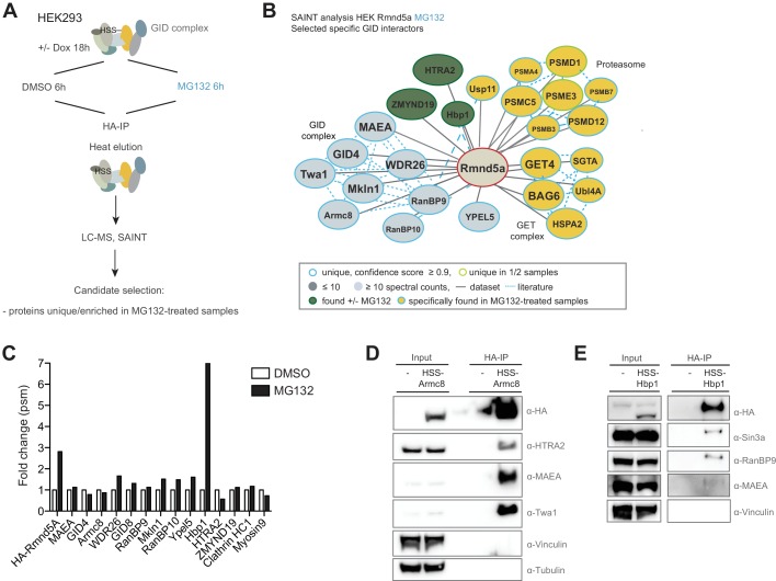
Figure 4—source data 1. List of Rmnd5a-interactors in the presence of MG132 identified by AP-MS and SAINT analysis.
elife-35528-fig4-data1.docx (138.7KB, docx)
DOI: 10.7554/eLife.35528.013
Figure 4—source data 2. List of Hbp1-interactors identified by AP-MS and SAINT analysis.
elife-35528-fig4-data2.docx (139.9KB, docx)
DOI: 10.7554/eLife.35528.014

Figure 4.

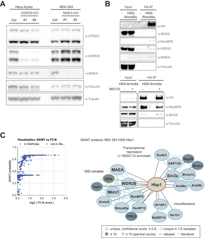
Figure 4—figure supplement 1. The transcription factor Hbp1 is a novel reciprocal GID-interacting protein.

Figure 4—figure supplement 1.

(A) The protein levels of the GID-interactor and mitochondrial protease HTRA2 was compared by immunoblotting of extracts prepared from HeLa Kyoto and HEK-293 cells deleted for WDR26 or MAEA using different gRNAs. (B) HA-immunoprecipitation of stably expressed HSS-Rmnd5a (upper panel) or HSS-Armc8 (lower panel) from HEK-293 cells. Where indicated (+), the cells were treated for 30 min with MG132 prior to cell lysis to prevent proteasomal degradation. Note that the mammalian GET complex protein BAG6 and the chaperone HSPA2 only weakly interact with the hGID complex, and the interaction of Rmnd5a with BAG6 is only marginally increased in the presence of MG132. (C) Graphical (left) and network (right) view of Hbp1-HCIPs mapped by AP-MS and subsequent SAINT analysis in HEK-293 cells (SAINT probability score ≥0.9, fold change over control (FC-B) ≥4, n = 2). Hbp1 shows two strong independent interaction modules comprising multiple subunits of the Sin3a co-repressor complex and the GID E3 ligase with all subunits present. Previously reported hits documented in the interaction database (iRefIndex) are displayed by red dots and novel hits are colored in blue.