. 2018 Jul 9;7:e35388. doi: 10.7554/eLife.35388

Figure 4. Engineering the Ire1-mediated non-conventional mRNA splicing in S. pombe cells.

(A) Measuring the non-conventional mRNA splicing in S. pombe cells, which were transformed with the S. cerevisiae HAC1 mRNA splicing reporter (SR) and the indicated Ire1 constructs. Cells were treated with 1 μg/ml Tm for 1 hr. (B) Illustration of the engineered S. pombe BIP1 mRNA splicing variant. (C) Measuring the non-conventional mRNA splicing of the engineered S. pombe BIP1 mRNA splicing variant. Experimental conditions are the same as those for Figure 4A. (D) Sequencing reads of the spliced BIP1 mRNA. The schematic illustration (E) and the splicing assay (F) of the synthetic splicing substrate in S. pombe. Cells were treated with 1 μg/ml Tm for 1 hr.

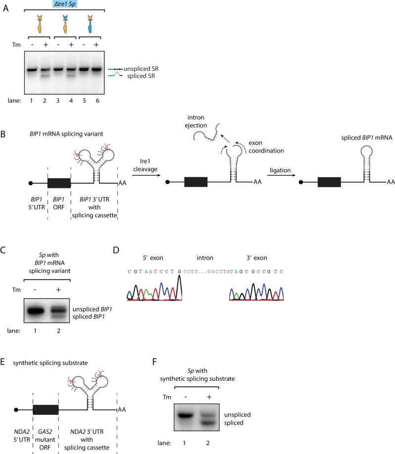
Figure 4.

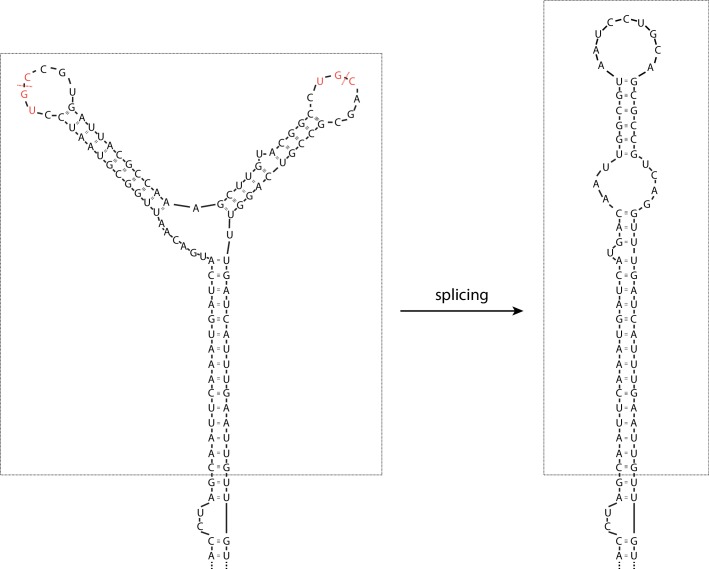
Figure 4—figure supplement 1. The splicing cassette in the engineered S. pombe BIP1 mRNA splicing variant.

Figure 4—figure supplement 1.

The part included in the dashed box is the inserted synthetic splicing cassette. The red dashed lines indicate the Ire1 cleavage sites. The S. pombe Ire1 UG|C motifs are labeled in red.
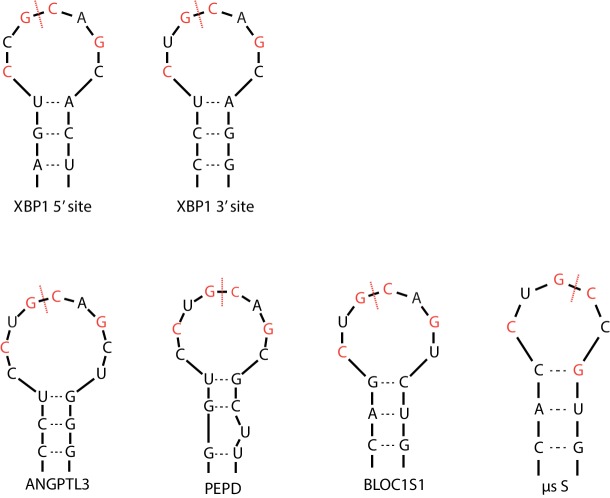
Figure 4—figure supplement 2. The Ire1α cleavage sites on XBP1 mRNA and RIDD targets.

Figure 4—figure supplement 2.

Red dashed lines mark the cleavage sites and the red letters indicate the previously identified sequence motif.
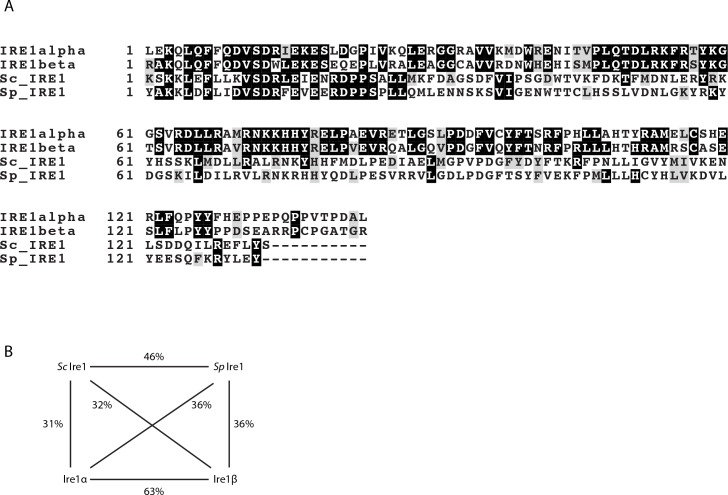
Figure 4—figure supplement 3. The sequence alignment of the kinase/RNase domains of Ire1α, Ire1β, the S. cerevisiae Ire1 and the S. pombe Ire1.

Figure 4—figure supplement 3.

(A) The sequence alignment and colored with BoxShade Server. (B) The sequence identities between the indicated pairs of Ire1 constructs.