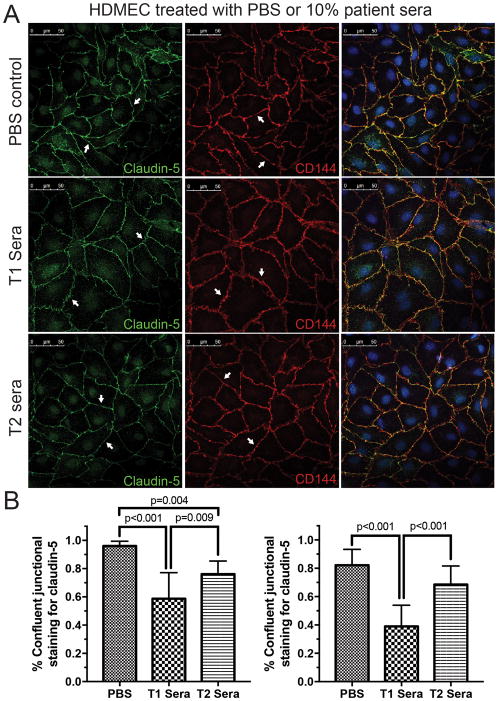
Figure 2.
Changes in the tight-junction protein claudin-5 and adherin-junction protein VE-cadherin in human dermal microvascular endothelial cells (HDMECs) after 6 hours of treatment with saline (PBS), pre- (T1) or post-CPB (T2) sera. (A) HDMECs display smooth and continuous junctional staining for both claudin-5 (Left panel) and VE-cadherin (CD144, Middle panel) in the untreated group (arrows). Contiguous junctional staining is disrupted and disjointed after 6 hours of stimulation with 10% pre-CPB sera (T1). The disruption of junctional staining is markedly less in cells treated with the immediately post-CPB serum (T2, arrows). Merged images indicate well defined junctional overlap (Right panels). Representative data from three experiments. (B) Quantification of circumferential disruption of junctional staining demonstrates significant differences in claudin-5 (left) and VE-cadherin (right) in HDMECs treated with PBS, T1 and T2 sera. Compiled data from three experiments, p values indicated.

