Fig. 3.

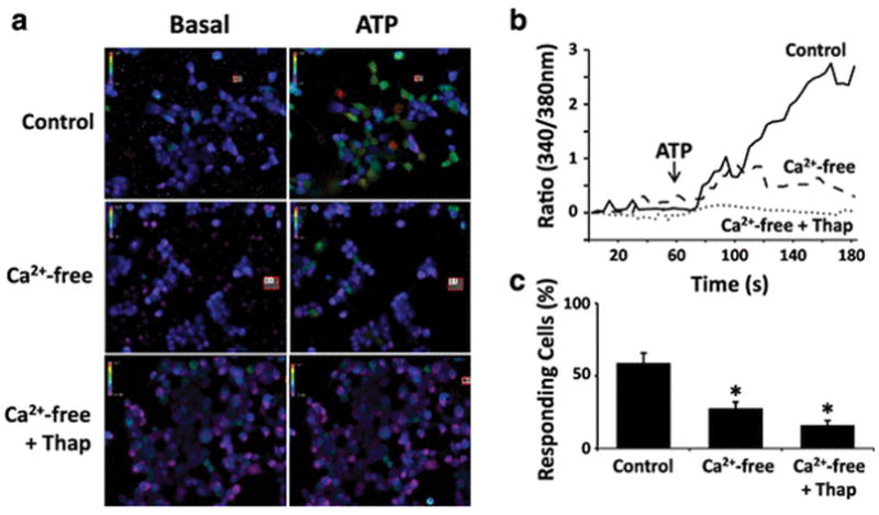
Calcium responses to ATP are attenuated in calcium-free solutions and after depletion of intracellular calcium stores. a Pseudocolored images depict intracellular calcium at basal levels and approximately 30 seconds following application of 200 μM ATP. In the experimental treatments, the extracellular solution contains physiological calcium (control), calcium-free solution, or calcium-free solution where intracellular calcium stores were first depleted by pre-treatment with 2 μM thapsigargin. b Graph depicting the average calcium responses of GL261 cells exposed to 200 μM ATP in control solution (solid line), calcium-free solution (dashed line), or calcium-free solution where intracellular calcium stores were first depleted by pre-treatment with 2 μM thapsigargin (dotted line). During analysis, 20 cells were selected for each experiment with no prior knowledge of the cells’ responsiveness to ATP. c Percentage of GL261 cells responding to ATP in control solution (n = 8), calcium-free solution (n = 16), and calcium-free solution where cells were first pre-treated with 2 μM thapsigargin (n = 26). Bars indicate standard error of the mean. Differences among groups were statistically significant (p < 0.01, one-way ANOVA). Pairwise comparisons between treatment groups were tested using the Holm-Sidak method; star (*) indicates groups that are significantly different from control
