Figure 1.
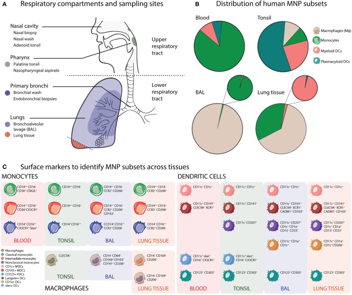
Mononuclear phagocyte (MNP) phenotype and distribution vary across human respiratory compartments. (A) Respiratory compartments and sampling sites. In the human upper respiratory tract, the initial site of influenza A virus infection, immune cells including macrophage (Mϕ), monocyte, and dendritic cell (DC) subsets from the nasal cavity and sinuses can be collected with nasal biopsies or nasal wash sampling. Along with pharyngeal palatine tonsils (and tubal and lingual tonsils), the adenoids form the Waldeyer’s ring, an anatomical structure comprising a ring of lymphoid tissue guarding the pharynx. In the lower respiratory tract, bronchoscopy allows sampling of discrete regions of the airways and lungs. Bronchial washes can be used to sample the cells lining the bronchi and bronchioles. Endobronchial biopsies can also be obtained from the mucosal tissue of the bronchial walls. Bronchoalveolar lavages (BALs) sample the most distal airways and alveolar sacs. Finally, lung resection samples allow sampling of lung parenchyma and tissue-resident immune cells. (B) Distribution of human MNP subsets. Pie charts illustrate broadly pooled data from 21 published studies on human MNP subset distribution in blood, tonsils, BAL, and lung tissue to demonstrate the differential distribution of MNPs across anatomical compartments reported from many research groups (51, 52, 57–61, 63–76). As different studies utilize different strategies to specifically define MNPs, the pie charts show groups of cells typically including several subsets of cells: Mϕs (beige), monocytes (green), myeloid DCs (MDCs) (coral), and plasmacytoid DCs (PDCs) (teal). (C) Surface markers to identify MNP subsets across human tissues. The various MNP subsets across tissues can be identified using flow cytometry from HLA-DR+ leukocytes that do not express lineage (T cells, B cells, NK cells, and granulocytes) markers. Apart from CD123+ PDCs, the MNP subsets express different levels of the myeloid marker CD11c. Mϕs have been studied in detail in both BAL and lung tissue, where CD169 expression distinguished alveolar from interstitial Mϕs. Monocyte subsets can be identified from most tissues based on relative expression of CD14 and CD16, as first defined in blood. The major MDC subsets are defined by expression of CD1c or CD141. The extended MDC subsets are now distinguished by expression of CD207 (langerin), CD1a, or slan (51, 52, 57–61, 63–76).

