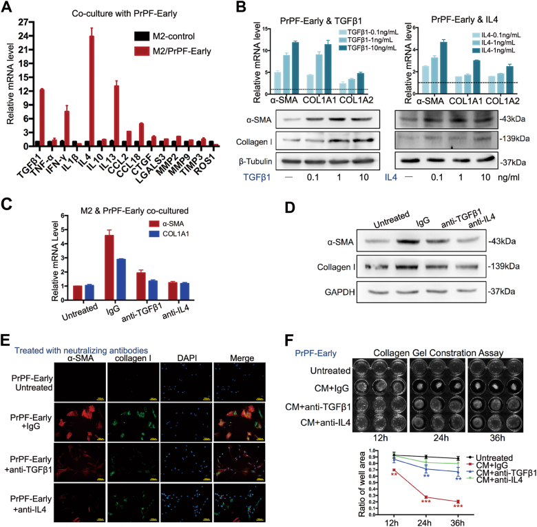
Fig. 3. TGFβ1 and IL4 may mediate the development of M2 macrophage-induced myofibroblast phenotype.
a Pro-fibrotic cytokine expression levels in the THP-1-derived M2 macrophages, co-cultured or not, with PrPF-early cells. b α-SMA, COL1A1 and COL3A1 expression, at mRNA and protein levels, following the exogenous addition of cytokines to PrPF-early samples compared to that in the untreated samples. The relative mRNA expression was compared with that in the untreated samples. c–f α-SMA and collagen I expression assessed using quantitative RT-PCR (c), western blotting (d), immunofluorescence staining (e) and contractility of PrPF (f) following the addition of anti-TGFβ1 (1 μg/ml), anti-IL4 (1 μg/ml) and IgG isotype (1 μg/ml) antibodies to the THP-1-derived M2 macrophage and PrPF-early cell co-cultures for 48 h, respectively. IgG isotype antibody was used as a control. Scale bar, 100 μm. All assays were performed in triplicate. *P < 0.05, **P < 0.01, ***P < 0.001

