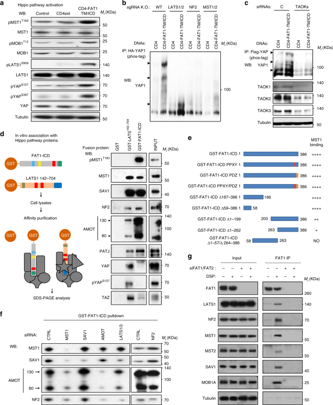
Fig. 3.
The intracellular domain of FAT1 interacts with and activates the Hippo kinase signalome. a Representative western blots against Hippo pathway components in lysates of exponentially growing HEK293 CD4ext and CD4-FAT1-TM/ICD stable cells. Control, parental HEK293 cell line. b Analysis of YAP1 phosphorylation after transient transfection with CD4 control or CD4-FAT1-TM/ICD chimera in WT or the corresponding CRISPR/Cas9 sgRNA engineered knockout HEK293 cells as indicated. HA-YAP1 immunoprecipitates were analyzed by phos-tag phosphorylation affinity shift electrophoresis and YAP1 western blotting. Retarded (phosphorylated) YAP1 is indicated by arrowheads. A representative blot is shown. c Analysis of YAP1 phosphorylation after transient cotransfection with Flag-YAP1 and CD4 control or CD4-FAT1-TM/ICD chimera in HEK293 cells pretreated with control (C) or TAOK1/2/3 siRNA (TAOKs) as indicated. Flag-YAP1 immunoprecipitates were analyzed by phos-tag electrophoresis and YAP1 western blotting. Retarded (phosphorylated) YAP1 is indicated by arrowheads. A representative blot is shown. d On the left, a scheme depicting the GST fusion proteins indicating the approximate location of functional motifs present in FAT1 and LATS1 and the subsequent GST-pulldown assay. On the right, representative western blots of pulldown experiments using GST fusion proteins and HEK293 total cell lysates. e Summary of mutant FAT1 ICD constructs (PPXY and PDZ binding site) and the depicted deletions and their ability to bind MST1 as assessed by pulldown assay. f siRNA-mediated knockdown on HEK293 of the different components of the Hippo signaling pathway and subsequent GST-FAT1-ICD pulldown on whole cells lysates. g Endogenous FAT1 immunoprecipitation by a monoclonal antibody recognizing is extracellular region. Exponentially growing HEK293 were transfected with FAT1 and FAT2 siRNAs for 48 h and then treated for 2 h at 4 °C with DMSO (−) or the reversible crosslinker DSP (+) prior to cell lysis and immunoprecipitation with anti-FAT1. Representative western blots are shown

