
Figure 1.
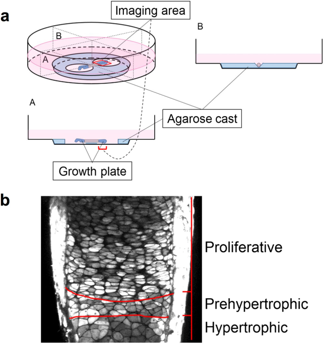
The dynamic imaging system of a fetal murine ulnar culture. (a) Setting of fetal murine ulnar culture. An explanted ulna was set on the agarose cast in the bottom of the glass-based dish, as shown in the schema. A red rectangle set on the distal growth plate, as indicated in the schema, represents an imaging area. The imaginary vertical-plane including longitudinal section of explanted ulna (A) and its maximal perpendicular vertical-plane (B) are indicated. (b) Definition of distinct areas of the growth plate in this imaging system. A captured photograph of the distal growth plate of explanted fetal murine ulna using two-photon excitation microscopy is shown. Red lines divide the growth plate into proliferative, prehypertrophic, and hypertrophic zones, as indicated.